All life is made up of cells a number of magnitudes smaller than a grain of salt. Their seemingly simple-looking buildings masks the intricate and sophisticated molecular exercise that allows them to hold out the capabilities that maintain life.
Researchers are starting to have the ability to visualize this exercise to a stage of element they have not been capable of earlier than.
Organic buildings might be visualized by both beginning on the stage of the entire organism and dealing down, or beginning on the stage of single atoms and dealing up.
Nonetheless, there was a decision hole between a cell’s smallest buildings, such because the cytoskeleton that helps the cell’s form, and its largest buildings, such because the ribosomes that make proteins in cells.
By analogy of Google Maps, whereas scientists have been capable of see whole cities and particular person homes, they didn’t have the instruments to see how the homes got here collectively to make up neighborhoods.
Seeing these neighborhood-level particulars is crucial to with the ability to perceive how particular person parts work collectively within the setting of a cell.
New instruments are steadily bridging this hole. And ongoing growth of 1 explicit method, cryo-electron tomography, or cryo-ET, has the potential to deepen how researchers examine and perceive how cells perform in well being and illness.
As the previous editor-in-chief of Science journal and as a researcher who has studied hard-to-visualize giant protein buildings for many years, I’ve witnessed astounding progress within the growth of instruments that may decide organic buildings intimately.
Simply because it turns into simpler to know how sophisticated programs work when you recognize what they seem like, understanding how organic buildings match collectively in a cell is essential to understanding how organisms perform.
A quick historical past of microscopy
Within the seventeenth century, gentle microscopy first revealed the existence of cells. Within the twentieth century, electron microscopy supplied even higher element, revealing the elaborate buildings inside cells, together with organelles just like the endoplasmic reticulum, a fancy community of membranes that play key roles in protein synthesis and transport.
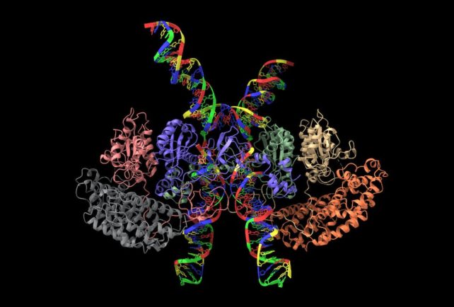
From the Nineteen Forties to Nineteen Sixties, biochemists labored to separate cells into their molecular parts and learn to decide the 3D buildings of proteins and different macromolecules at or close to atomic decision. This was first completed utilizing X-ray crystallography to visualise the construction of myoglobin, a protein that provides oxygen to muscle groups.
Over the previous decade, strategies primarily based on nuclear magnetic resonance, which produces photos primarily based on how atoms work together in a magnetic area, and cryo-electron microscopy have quickly elevated the quantity and complexity of the buildings scientists can visualize.
frameborder=”0″ enable=”accelerometer; autoplay; clipboard-write; encrypted-media; gyroscope; picture-in-picture; web-share” allowfullscreen>
What’s cryo-EM and cryo-ET?
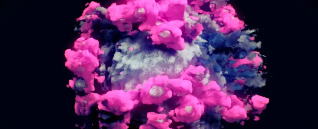
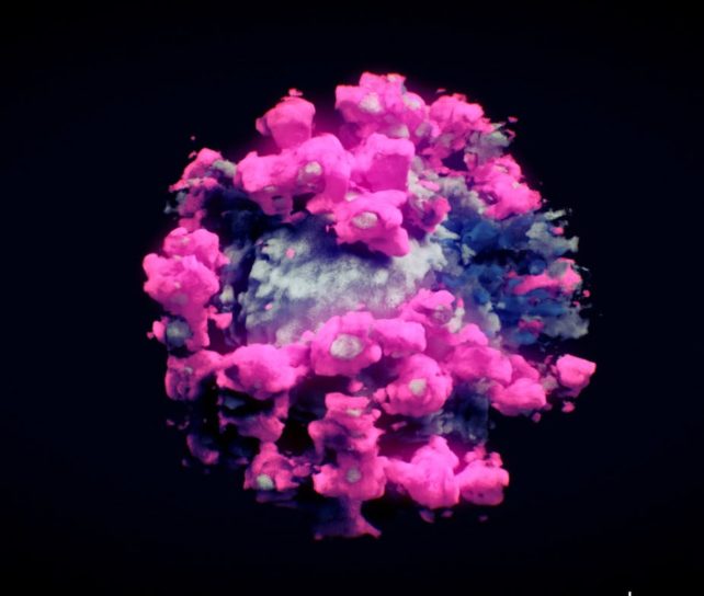
Cryo-electron microscopy, or cryo-EM, makes use of a digicam to detect how a beam of electrons is deflected because the electrons move by means of a pattern to visualise buildings on the molecular stage.
Samples are quickly frozen to guard them from radiation injury. Detailed fashions of the construction of curiosity are made by taking a number of photos of particular person molecules and averaging them right into a 3D construction.
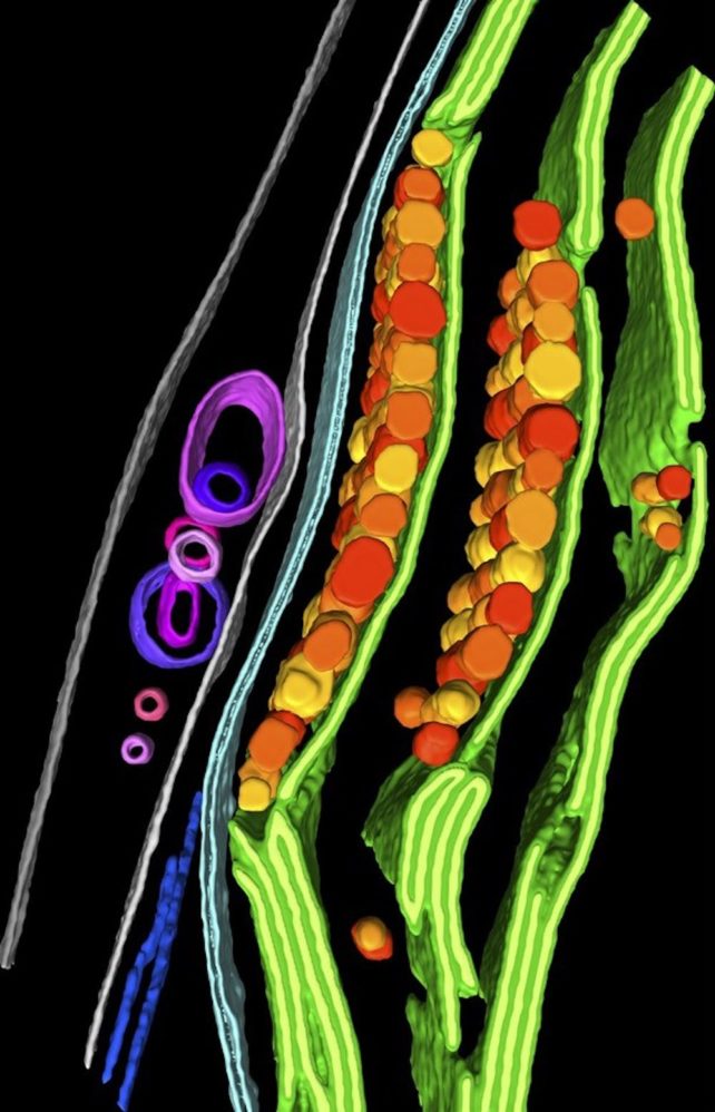
Cryo-ET shares comparable parts with cryo-EM however makes use of totally different strategies. As a result of most cells are too thick to be imaged clearly, a area of curiosity in a cell is first thinned through the use of an ion beam.
The pattern is then tilted to take a number of footage of it at totally different angles, analogous to a CT scan of a physique half – though on this case the imaging system itself is tilted, quite than the affected person. These photos are then mixed by a pc to provide a 3D picture of a portion of the cell.
The decision of this picture is excessive sufficient that researchers – or pc applications – can determine the person parts of various buildings in a cell. Researchers have used this method, for instance, to point out how proteins transfer and are degraded inside an algal cell.
Most of the steps researchers as soon as needed to do manually to find out the buildings of cells have gotten automated, permitting scientists to determine new buildings at vastly larger speeds.
For instance, combining cryo-EM with synthetic intelligence applications like AlphaFold can facilitate picture interpretation by predicting protein buildings that haven’t but been characterised.
Understanding cell construction and performance
As imaging strategies and workflows enhance, researchers will be capable to sort out some key questions in cell biology with totally different methods.
Step one is to determine what cells and which areas inside these cells to check. One other visualization method known as correlated gentle and electron microscopy, or CLEM, makes use of fluorescent tags to assist find areas the place fascinating processes are going down in residing cells.
Evaluating the genetic distinction between cells can present further perception. Scientists can have a look at cells which might be unable to hold out explicit capabilities and see how that is mirrored of their construction. This method may assist researchers examine how cells work together with one another.
Cryo-ET is prone to stay a specialised software for a while. However additional technological developments and rising accessibility will enable the scientific group to look at the hyperlink between mobile construction and performance at beforehand inaccessible ranges of element.
I anticipate seeing new theories on how we perceive cells, transferring from disorganized luggage of molecules to intricately organized and dynamic programs.![]()
Jeremy Berg, Professor of Computational and Techniques Biology, Affiliate Senior Vice Chancellor for Science Technique and Planning, College of Pittsburgh
This text is republished from The Dialog underneath a Inventive Commons license. Learn the unique article.