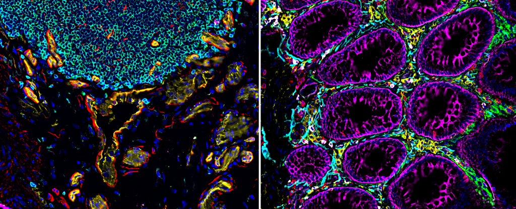
A consortium of scientists has simply printed an atlas of outstanding photographs of three human organs, every important in their very own manner, displaying how cell varieties are organized and work together.
The consequence: Glittering, kaleidoscopic blueprints lit up by fluorescent dyes that reveal new intimacies about our our bodies and reshape our understanding of human biology and illness like by no means earlier than.
As you’ll be able to see within the diagram under, researchers generated the cell atlases in 3 ways.
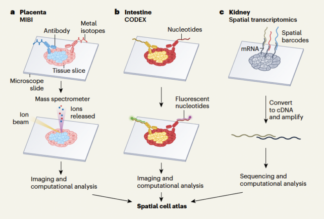
One crew, led by Washington University nephrologist Sanjay Jain, used single-cell transcriptomics strategies, which reveal how the genetic directions encoded in DNA are learn in particular person cells, to map the kidney.
Another group headed up by genomicist Michael Snyder at Stanford School of Medicine mapped the gut with fluorescent antibodies certain to tissue sections, imaged beneath the microscope.
And a 3rd crew peered into what scientists have described as “arguably crucial organ of the physique, however paradoxically essentially the most poorly understood” – the placenta.
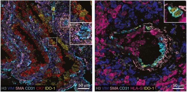
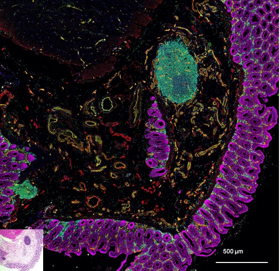
Stanford University pathologist Michael Angelo and colleagues imaged slices of placental tissue handled with steel ions chemically linked to antibodies able to latching onto signature compounds on the surfaces of cells, specializing in samples the place placental cells had hooked up to the wall of the uterus.
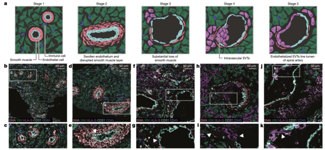
By imaging a number of samples at totally different levels of this course of, from 6 to twenty weeks’ gestation, researchers plotted interactions between placental cells and the mom’s immune cells and arteries – each of which modify to accommodate the placenta.
We can see, in beautiful element, how this reworking course of “permits peaceable coexistence between genetically distinct maternal uterine and fetal placental cells,” in response to two cell biologists on the Wellcome Sanger Institute, Roser Vento-Tormo and Roser Vilarrasa-Blasi, who penned a commentary concerning the assortment of latest papers.
As for the gut, this meters-long organ is the place thousands and thousands of microbes jostle about, ultra-processed meals set off irritation, and cells are plugged into the physique’s ‘second mind‘.
Snyder’s crew found drastic shifts in how cells have been organized alongside the size of the gut. They sketched out distinct neighborhoods stacked with immune cells able to launch into motion and stumbled upon new subtypes of epithelial cells that line the intestine.
Further imaging of the ruffled surfaced of the gut and its layers might reveal new insights about how inflammatory bowel ailments, temper issues, and even neurodegenerative ailments develop.
Kidneys, too, achieve this a lot for the physique. They pump blood to scrub it of poisons and waste merchandise, however usually fail or develop into diseased and want changing.
Sampling greater than 90 kidneys, Jain and colleagues outlined communication channels between cells and situated cells by which restore pathways develop into faulty throughout acute kidney harm or persistent kidney illness.
“We checked out how kidney cells are organized, their molecular identities, and the way they shift from wholesome to diseased states,” explains Jain, who led the kidney imaging research.
“With this information, we are able to begin excited about the medicine or small molecule targets that may forestall development to illness or promote restoration from harm.”
Bear in thoughts these photographs have been taken utilizing a small variety of valuable samples generously donated by folks present process surgical procedure and volunteering to take part in analysis.
A a lot more durable job will likely be appreciating the structural variations in organs that exist between totally different teams and populations that may have actual well being penalties, even once we’re all made to roughly the identical physique plan.
The papers and the accompanying commentary have been printed collectively in Nature.